Chromosome Structure/Centromere/Telomere

Chromosomes have essential parts called centromeres and telomeres. These are crucial regions on chromosomes necessary for the accurate transmission of DNA from parent to offspring and from cell to cell.
Table of Contents
Chromosome Structure
Chromosomes contain parts known as centromeres and telomeres. These are critical regions on chromosomes necessary for the accurate transmission of DNA from parent to offspring and from cell to cell.
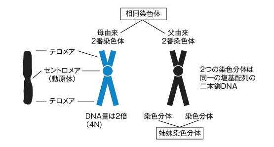
The central part where two chromosomes are attached is called the centromere.
From the centromere, it appears that arms are extending. The shorter arm is called the short arm (p) and the longer arm is called the long arm (q). The bridge between each arm is called the telomere.
Chromosomes during division are replicas of DNA made in the same way.
One of these is called a chromatid, and two together are called sister chromatids.
Although these sister chromatids are made from identical DNA, cells originally contain pairs of chromosome DNA. These are called homologous chromosomes when they become chromosomes, 1 through 22 are called autosomes, and X and Y are called sex chromosomes.
Role of Centromere
Centromeres and kinetochores were once thought to be the same, but kinetochores often refer to the chromosomal (DNA) region associated with kinetochores, and kinetochores often refer to the attachment structure itself, known as microtubules.
The centromere has two roles in meiosis.
The first is that it is a region that accurately distributes chromosomal DNA to two daughter cells.
The second is that it is controlled to separate sister chromatids accurately.
The telomere is a complex of special structures and proteins at the ends of one of the two strands of double-stranded DNA that make up a chromosome. The telomere tends to shorten with age.
X Chromosome Inactivation
Women have two X chromosomes, and men have one.
There is something called X-chromosome inactivation. Men have one small Y chromosome in addition to their X chromosome, but women have two X chromosomes, resulting in an excessive amount of X-chromosome genes.
Therefore, to equalize gene expression on X chromosomes between males and females, a mechanism exists to inactivate one of the two X chromosomes and suppress gene activity. This X-chromosome inactivation is also known as Lyonization. Inactivation on the X chromosome occurs randomly regardless of maternal or paternal origin. Once the X chromosome is inactivated, it remains inactive in subsequent daughter cells. This is inherited through mitosis.
Inactivation when there are more than two X chromosomes
So, what happens if there are three or four X chromosomes? This can be examined using a test called FISH.
If there are three X chromosomes, two out of the three will be inactivated. If there are four X chromosomes, three out of the four will be inactivated. One X chromosome is always active. If this inactivation did not occur, what would happen? Inactivation is necessary to regulate gene dosage, preventing severe developmental delays due to excessive gene dosage. In most cases, it is believed this would result in miscarriage.
Also, if there are three X chromosomes and only very small fragments of the third X chromosome remain active, severe developmental delays occur from birth. Additionally, occasionally there is a mixture of cells in which the maternal X chromosome is inactivated and cells in which the paternal X chromosome is inactivated. This is called mosaicism.
Recently, we often hear about ES cells and iPS cells, which have not undergone inactivation themselves, and as cells differentiate, one or the other is inactivated.