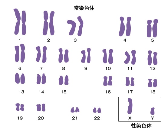
The Number and Morphology of Human Chromosomes

Human chromosome analysis is referred to as cytogenetics, which encompasses the genetic realm of chromosomes.
It is a composite term combining cytology (the study of cells) and genetics.
Table of Contents
Genetics of Chromosomes
What is Human Chromosome Analysis?
The genetic realm of chromosomes is referred to as cytogenetics, a composite term combining cytology and genetics.
Mendel's "laws of inheritance" in pea plants were rediscovered in 1900, revealing a consistent pattern of heredity from parent to offspring through meiosis, closely linked with chromosomes. Subsequently, Morgan elucidated the relationship between genes and chromosomes using Drosophila.
Detailed observation and analysis of human chromosome number and morphology have not been possible for long.
In 1956, Théo and Lejeune revealed using cultured fetal cells that humans have 46 chromosomes (2n=46), with males having XY and females having XX sex chromosomes.
Since then, advancements in cell culture and chromosome specimen preparation have enabled the identification of various chromosomal abnormalities related to congenital conditions and hematologic disorders. The precision of chromosome analysis has advanced significantly, leading to the establishment of classification and nomenclature standards for human chromosomes.
Since 1960, standard conventions for chromosomes have undergone multiple revisions.
Technological innovations in chromosome analysis around 1970, such as banding techniques, enabled the identification of all human chromosomes and accurately pinpointed structural abnormalities using banding patterns and symbols.
Analysis of Chromosomal Structural Abnormalities
By the 1980s, highly accurate banding techniques were developed, enabling detailed analysis of chromosomal structural abnormalities.
Around the 1990s, molecular genetic methods were introduced into the field of cytogenetics, allowing chromosome analysis at the DNA level. During this time, the Human Genome Project was initiated with the aim of sequencing the entire genome's base pairs, unraveling genetic information and its regulatory mechanisms.
Entering the 2000s, advancements in human genome analysis led to the development of high-resolution chromosome DNA analysis using microarrays, leveraging databases containing comprehensive DNA sequence information. This enabled the detection of partial deletions, duplications on chromosome DNA, and even continuous counts of DNA base pairs.
Human Chromosome Map
In 1990, the international Human Genome Project was launched with the goal of sequencing the entire genome's base pairs to unravel the complete picture of genetic information and its regulatory mechanisms.
The sequencing of small, fragmented DNA clones accelerated rapidly, and by 2003, the complete genome sequence was reported.
In human genome analysis, the number of genes is estimated to be between 22,000 to 25,000.
In mammals, including mice, both the genome size and the number of genes are comparable to humans. The human genome is approximately 3,100 Mb (3.1 billion base pairs).
The average size of gene regions, which include exons and introns, is about 33 kb. Therefore, genes occupy less than 30% of the human genome (3,100 Mb). The majority of the genome consists of regions other than genes (as mentioned earlier).
Genes: Their Positions and Numbers
Genes are not evenly distributed along DNA molecules; rather, DNA is tightly folded into chromosomes in a highly organized manner. Specific genes are located at defined positions on chromosomes.
These bands on chromosomes exhibit regions of varying gene density along the vertical axis. The uneven distribution of genes across the genome can be observed on chromosomes, indicating the number of genes present on each chromosome.
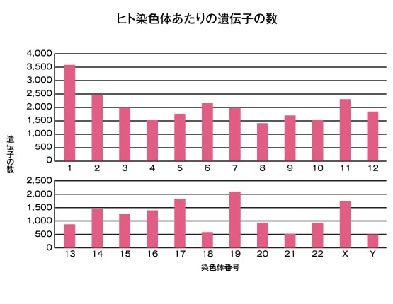
The number of genes is not directly correlated with the size of chromosomes. For instance, smaller chromosomes like chromosome 19 can have a much higher number of genes compared to other chromosomes of similar size.
Additionally, trisomies that are viable at birth, such as trisomies of chromosome 13, 18, and 21, have fewer genes compared to other chromosomes.
Because these chromosomes naturally have fewer genes, they can still result in viable births even when an extra copy (trisomy) is present. In contrast, trisomies involving other chromosomes typically lead to intrauterine growth arrest and miscarriage due to the greater genetic imbalance.